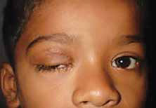
Traumatic Ptosis: Which one has the best prognosis?
Jacons SM et al investigated whether a systematic approach to subgrouping traumatic ptosis according to etiology can allow for better tailoring of prognosis and treatment. A retrospective chart review of patients with traumarelated blepharoptosis managed by oculoplastic surgery specialists at an academic medical center from January 1995 to November 2015 was done. Injury mechanism, eyelid position and function, interventions, and outcomes were reviewed. Of 648 patients treated for blepharoptosis, 55 (8.5%) were traumatic. Careful review revealed 4 subcategories of traumatic ptosis cases: aponeurotic (n = 16), myogenic (n = 18), neurogenic (n = 7), and mechanical (n = 14). Margin reflex distance (MRD1) at presentation was significantly worse for the myogenic subtype (-0.59 mm, SD ±2.09, P = 0.046). The aponeurotic subtype had the best average levator function at presentation (14.29 mm, SD ±2.05), while myogenic had the worst (8.41 mm, SD ±4.94) (P = 0.004). Thirty-five (63.6%) patients were managed surgically. Final MRD1 was significantly different for each subtype (P = 0.163), with aponeurotic 2.63 mm (SD ±1.01), myogenic 1.29 mm (SD ±2.24), neurogenic 1.79 mm (SD ±2.48), and mechanical 2.31 mm (SD ±1.18). There was a significant increase in MRD1 from presentation to final follow up across all groups (P < 0.05). The authors concluded that traumatic ptosis is fairly heterogenous. Systematically evaluating traumatic ptosis cases by trauma mechanism can guide decisions about prognosis and management. Two-thirds of cases require surgical treatment, with most patients responding well to conjunctiva-Müller resection or external levator advancement. While all subgroups demonstrated improvement in MRD1 at final follow up, aponeurotic cases had the best prognosis, while myogenic fared the worst and required the longest for maximal recovery.
Read More:
Jacobs SM et al. Traumatic Ptosis: Evaluation of Etiology, Management and Prognosis. J Ophthalmic Vis Res. 2018 Oct-Dec;13(4):447-452.
Eyelid pressure causing ocular surface disorders
Yamaguchi et al hypothesised that excessive pressure by the eyelid onto the corneal surface can result in development or worsening of ocular surface disorder. To prove this they took the help of an interesting device called blepharotensiometer which can determine changes in eyelid pressures. They evaluated various types of ocular surface disorder patients with this device. The methodology involved four steps. First, the repeatability of the blepharo-tensiometer was determined by measuring the eyelid pressures on 3 separate days from healthy volunteers and calculating the intraclass correlation coefficients (ICCs). Second, to determine the ability of the blepharo-tensiometer to detect changes in the eyelid pressures in different types of ocular surface disorders, we compared the eyelid pressure of healthy eyes with dry eyes. Third, the correlations between the eyelid pressure and the location and magnitude of fluorescein staining of the ocular surface were analyzed. Fourth, the eyelid pressure in eyes with lid-wiper epitheliopathy (LWE) was measured. The ICCs ranged from 0.675 to 0.911 for the upper eyelid and 0.663 to 0.925 for the lower eyelid. The pressures of the upper and lower eyelids were significantly higher in dry eyes than in healthy eyes. The inferior ocular surface staining scores were strongly correlated with the lower eyelid pressure by multivariate analysis. The lower eyelid pressure was significantly correlated with the grade of the lower LWE. The study found that the blepahro-tensiometer can obtain repeatable measurements of the eyelid pressure and can be used to evaluate the pressure of the eyelids on the ocular surface in healthy and diseased eyes. The authors concluded that significant correlations between the eyelid pressure and the ocular surface staining suggests that the pressure on the ocular surface probably contributes to ocular surface disorders
Read More:
Yamaguchi M, Shiraishi A. Relationship Between Eyelid Pressure and Ocular Surface Disorders in Patients With Healthy and Dry Eyes. Invest Ophthalmol Vis Sci. 2018 Nov 1;59(14).