
Daraius Shroff, MS FRCS
Vitreoretinal and Imaging Services
Shroff Eye Centre, New Delhi

Priyanka Gupta, DNB
Vitreoretinal and Imaging Services
Shroff Eye Centre, New Delhi

Charu Gupta, MS
Vitreoretinal and Imaging Services
Shroff Eye Centre, New Delhi

Anuj Choudhary, B.Opt
Vitreoretinal and Imaging Services
Shroff Eye Centre, New Delhi

Cyrus M Shroff, MD
Vitreoretinal and Imaging Services
Shroff Eye Centre, New Delhi
Retinal diseases are a major cause of impaired vision especially in the ageing population. It is therefore not surprising that advances in retinal imaging and the treatment of retinal diseases are an active area of research. Early recognition of pathology permits timely management of retinal diseases. Year after year, technical advances have enabled us to image wider and deeper and less invasively. These imaging systems are helpful in diagnosis, treatment and monitoring of disease progression or response to therapy in patients with retinal disorders.
In this article, we discuss how newer technological advances in imaging are improving our management of retinal diseases.
Targeted retinal photocoagulation and laser therapy based on ultrawide field imaging
Current generation of ultra-widefield imaging permits acquisition of 2000 of the fundus in a single image. Areas of capillary non-perfusion or neovascularization in extreme peripheries, which could be missed on conventional imaging systems, can be easily detected. The relative position of the lesions with respect to other structures need not be extrapolated and multiple frames are not needed for correlation or to see extent of pathology with this new imaging technique.
This leads to focussed delivery of treatment to the area of pathology.1,2,3 Areas of peripheral capillary non perfusion (Figure 1a), which would be missed on conventional imaging systems can be picked up, leading to their targeted treatment (Figure 1b) and avoiding treating healthy retina.4 Even far peripheral neovascularisation can be picked up and accurately treated in a targeted fashionon ultra-wide field angiography (Figure 1c).
Non-invasive imaging with OCT Angiography (OCTA) and its practical uses
OCT angiography (OCTA) is a non-invasive three dimensional imaging technique for visualizing the retinal and choroidal vasculature providing depth – resolved structural and functional information regarding blood flow in these vessels.5,6 OCTA in diagnosis and subsequent treatment OCTA helps in delineating vascular networks even in clinical situations where fundus fluorescein angiography (FFA) and indocyanine green (ICG) angiography may not be informative. The usefulness OCTA is well exemplified in a 64-year-old gentleman who received multiple ranibizumab injections for a provisional diagnosis of subretinal fluid (SRF) with suspected CNVM. FFA (Figure 2a and b) and ICG (Figure 2c and d) was done. However, the diagnosis could not be ascertained until OCTA revealed a classic vascular network (Figure 3a and b). Enhanced depth imaging (EDI) confirmed thickened choroid suggestive of pachychoroid spectrum. (Figure3c). Subsequent treatment with aflibercept resulted in resolution of fluid and improvement in vision.
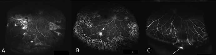
Figure 1: Panel A Shows Ultra- wide field FFA
showing non perfusion areas prior to treatment.Panel B shows ultra-wide field FFA following targeted laser photo coagulation. Panel C shows ultra- wide field FFA pointing far peripheral secularization at the pars plana.

Figure 2: Patient with doubtful CNVM
Panel A and B – FFA (arteriovenous phase) show stippled hyperfluoroscence in both eyes. Panel C and D – ICG angiography shows dilated choroidal vessels in both eyes but no definite CNVM can be delineated.
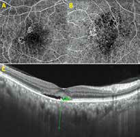
Figure 3: Patient with doubtful CNVM
Panel A shows small vascular network suggesting CNVM temporal to fovea in the right eye. Panel B shows the vascular network with central void area in the left eye. Panel C – EDI OCT shows increased choroidal thickness with persistence of SRF in the right eye suggestive of Pachychoroid.
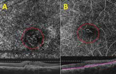
Figure 4: OCTA image (panel A) shows small vascular network depicting CNVM
(red circle) Pre Anti VEGF therapy. Corresponding B scan OCT shows CNV with small cystic space and. Panel B shows regression of network following anti- VEGF therapy. Corresponding B scan OCT also shows regression of CNV with resolution of cystic space.
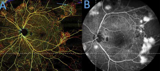
Figure 5: Montage OCTA image (panel A) showing gross capillary non perfusion
in all quadrants with active NVE (arrow). Panel B showing FFA showing leaking NVE and non-perfusion.
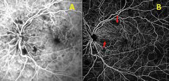
Figure 6: Central retinal vein occlusion as seen SLO based FFA
(panel A), shows ischemic area with blurred boundaries. compared to OCTA (panel B) image showing classic ischemic areas with capillary changes which are clearly delineated (arrow)
informative. The usefulness of OCTA is well exemplified in a 64-year-old gentleman who received multiple ranibizumab injections for a provisional diagnosis of subretinal fluid (SRF) with suspected CNVM. FFA (Figure 2a and b) and ICG (Figure 2c and d) was done. However, the diagnosis could not be ascertained until OCTA revealed a classic vascular network (Figure 3a and b). Enhanced depth imaging (EDI) confirmed thickened choroid suggestive of pachychoroid spectrum. (Figure 3c). Subsequent treatment with aflibercept resulted in resolution of fluid and improvement in vision.
OCTA in follow up and monitoring of therapy
OCTA allows study & classification of new vessels, highlighting their morphology6,7. By provide quantitative information regarding choroidal neovascularisation (CNV) flow, vessel density and lesion area (Figure 4a), OCTA can be used as a tool for monitoring the response to anti-VEGF therapy (Figure 4b).
Wide field imaging with OCTA
Montage images Swept source OCTA is a new addition to the imaging armamentarium. By using this advanced technology we are able to accurately put together images from different quadrants. This is done by an inbuilt software thereby providing a beautiful montage view (Figure 5a & 5b) with a much larger field of view. This enables us to image areas of neovascularization as well as capillary non perfusion non-invasively. It is also possible to analyse vascular changes at varying depths.
OCTA for vascular occlusion
Characteristic findings of acute and chronic retinal vein occlusions are well demonstrated on OCTA. The superficial and deep capillary plexus are easily differentiated on OCTA which are otherwise obscured by leakage of dye in FFA. The vessels in the superficial plexus close to the foveal avascular zone (FAZ) are narrower and more tortuous. Loss of
capillary perfusion is usually more marked in the deep plexus which is better appreciated on montage view (Figure 6a and b).
Conclusion
Improved retinal imaging due to new technological advances has helped us in the accurate diagnosis and localization of a variety of pathologies. This in turn favourably alters our management and clinical outcomes of the treatment of retinal diseases.
References:
1. Wessel et al. Ultra-wide-field angiography improves the detection and classification of diabetic retinopathy. Retina
2012;32:785–791.
2. Kang KB et al. Ultra- widefield imaging for the management of pediatric retinal diseases. J Pediatr Ophthalmol Strabismus. 2013 Sep-Oct:50(5):282-8.
3. Shroff D, Narain S, Gupta C, Dutta R, Shroff C. Non-contact Ultra-widefield Imaging in Lasered Retinopathy of
prematurity. Indian J Pediatr. 2015 Dec 17.
4. Ultra Wide Field Fluorescein Angiography Guided Targeted Retinal Photocoagulation (TRP). Reddy S, Hu A, Schwartz SD. Semin Ophthalmol. 2009;24(1):9-14.
5. de Carlo, Romano A, Waheed NK, Duker JS. A review of optical coherence tomography angiography(OCTA). International journal of Retina and Viterous. 2015 Feb1:5.
6. F. Bandello. OCT Angiography in Retinal and Macular Diseases. Dev Ophthalmol. Basel, Karger,2016, vol 56.
7. Coscas et al. OCT Angiography in exudative AMD. Retina; 2015; 35: 2219-28.