
Renuka Raghvendra Chakurkar, DNB
L.V.Prasad Eye Institute, Hyderabad

Jay Chhablani, MS
L.V.Prasad Eye Institute, Hyderabad
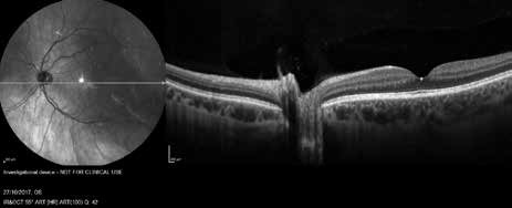
Figure 1: Wide field OCT image of a normal eye
(acknowledgment – Dr. Marco Lupidi).
Advances in retinal imaging have led to change in practice patterns in vitreo-retina subspeciality. This article highlights emerging imaging techniques, which has the potential to further renovate the management of retinal diseases.
Multicolour Imaging
In multicolour imaging, the retina and optic nerve are scanned simultaneously with three lasers of different wavelengths: infrared (815 nm), green (518 nm), and blue (486 nm).(1) The shorter wavelength blue laser provides details of superficial retinal structures such as retinal nerve fiber layer, ganglion cells, macular pigment, and any structures on the surface of the retina like epiretinal membranes. The medium wavelength green laser is mainly absorbed by hemoglobin and provides details of vascular abnormalities such as microaneurysms. The longer wavelength infrared laser penetrates the deepest – providing images of the retinal pigment epithelium (RPE) and the choroid and is particularly useful for imaging deeper lesions such as choroidal rupture and nevi.
Multicolour imaging is obtained by using multiple laser wavelengths simultaneously to selectively capture different retinal structures from a single image during a single examination. It helps in easier and earlier detection of various pathologies such as agerelated macular degeneration and diabetic retinopathy as compared to a conventional colour photograph
Wide field OCT
Wide field OCT system can obtain 70,000 A-scans/second and provides 12 mm x 9 mm B-scans, in contrast to 6 to 9 mm scan length of available OCT systems. It provides detailed imaging of both the choroid and the retina with active eye tracking enhancing image stability.
Wide field OCT enhances visualization of macular disorders that span greater areas than standard scan widths eg. multifocal central serous retinopathy.(2) It also expands our understanding of peripheral vitreoretinal diseases and choroidal lesions (eg, malignancies)
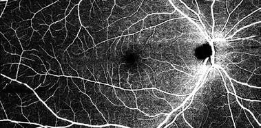
Figure 2: Wide field OCTA image of a normal eye
(acknowledgment – Dr. Marco Lupidi)
Wide field OCTA
The main limitations of OCTA are limited field of view and eye motion artefacts.(3) The wide-field OCTA provides the depth-resolved information and detailed visualization of vascular network upto 12 X 12 mm2 area which is very useful in patients with venous occlusion and DR patients, is showing ischemic areas and hidden neovascularization without any need for invasive angiography
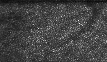
Figure 3: Adaptive optics images of a normal
eye showing the rods and cones
(Dark and Bright Dots).
Spectral Imaging
Spectroscopy is the study of the interaction between any form of matter and radiated energy. The combination of spectroscopy with conventional imaging techniques – “spectral imaging” – allows determination of the spatial distribution of spectroscopic data. (4) The spectroscopic principles have been used for oximetry, the measurement of oxygen saturation in a patient’s blood.
Spectral imaging devices typically employ one of three approaches:
(1) multispectral imaging.
(2) hyperspectral imaging.
(3) imaging spectroscopy. This technique is dependent on assumptions about the relationship between light transmittance through the blood and its oxygen saturation (according to the Lambert-Beer law).
Knowledge about the oxygen saturation is important in retinal vascular occlusions and diabetic retinopathy.(5, 6) Its application has been shown in eyes with glaucoma, as well in retinal dystrophies.(7) Future prospects for oximetry include integration with a scanning laser ophthalmoscope and its application in predicting systemic disease from retinal oxygen saturation values.
Adaptive Optics
The aberrations in the optical system of eye reduce the lateral resolution of retinal images by OCT. The adaptive optics devices use wavefront sensing to compensate for such aberrations. Adaptive optics components have been incorporated into confocal SLO systems with the advantages of increased contrast, cross-sectional imaging, and the
measurement of dynamic changes such as blood flow. (8)
The adaptive optics retinal imaging allows direct measurement of photoreceptor density and diameter (Figure 4) and is useful for the examination of patients with inherited retinal degenerations.(9) Limited field of imaging, long image acquisition time, unavailability of validated automated algorithms are the major limitations.
References:
1. Sergott RC. Retinal segmentation using multicolor laser imaging. Journal of Neuro-Ophthalmology. 2014;34:S24-S8.
2. LEVISON A. Widefield OCT: Current and Future Applications.
3. Klein T, Wieser W, Eigenwillig CM, Biedermann BR, Huber R. Megahertz OCT for ultrawidefield ultrawidefield retinal imaging with a 1050nm Fourier domain modelocked laser. Optics express. 2011;19(4):3044-62.
4. Mordant D, Al-Abboud I, Muyo G, Gorman A, Harvey A, McNaught A. Oxygen saturation measurements of the retinal vasculature in treated asymmetrical primary open-angle glaucoma using hyperspectral imaging. Eye. 2014;28(10):1190.
5. Hardarson SH, Stefánsson E. Retinal oxygen saturation is altered in diabetic retinopathy. British journal of ophthalmology. 2012;96(4):560-3.
6. Hardarson SH, Stefánsson E. Oxygen saturation in branch retinal vein occlusion. Acta Ophthalmologica. 2012;90(5):466-70.
7. Eysteinsson T, Hardarson SH, Bragason D, Stefánsson E. Retinal vessel oxygen saturation and vessel diameter in retinitis pigmentosa. Acta ophthalmologica. 2014;92(5):449-53.
8. Liang J, Williams DR, Miller DT. Supernormal vision and high-resolution retinal imaging through adaptive optics. JOSA A. 1997;14(11):2884-92.
9. Wolfing JI, Chung M, Carroll J, Roorda A, Williams DR. High-resolution retinal imaging of cone–rod dystrophy. Ophthalmology. 2006;113(6):1014-9. e1.